Micro X-ray Computed Tomography (µ-CT)
This facility was established in May 2017 with the purchase of the Bruker SkyScan 1276, an in vivo X-ray microtomograph. Our µ-CT offers high spatial resolution, the ability to differentiate between different tissue densities and it allows three-dimensional visual reconstructions of tissue. The SkyScan 1276 is suitable for analysis of bones, vasculature, lung, heart, body fat and tumors of living animals like rat, mice and smaller species, but also qualifies for ex-vivo analyses.
Below, images of selective projects on various species and organs are presented.

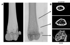
Fig. 8: Microcomputer tomography (µCT) of mouse distal femur. (A) longitudinal view of distal femur (B) transaxial view from distal to proximal; bottom: transaxial view of the epiphyseal reagion of the femur; middle: trabecular bone, transaxial view of the metaphyseal region; top: transaxial view of diaphyseal region. Scan options: X-ray voltage: 70 KV, X-ray current: 200 µA, filter: Al 0.5 mm, image pixel size: 4 µm, tomographic rotation: 180°, rotation step: 0.200°, frame averaging: 8, scan duration: 1h15min. Pictures provided by Sabrina Sapski, Department II.

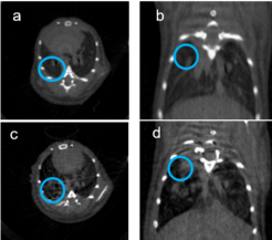
Fig. 9: In-vivo microcomputer tomography (µCT) of mouse lung. (a,b) show the mouse lung prior tumor cell inoculation and (c,d) the same mouse lung two weeks post intratracheal injection of Lewis Lung Carcioma cells. Scan options: X-ray voltage: 50 KV, X-ray current: 200 µA, filter: Al 0.5 mm, image pixel size: 84.8 µm, tomographic rotation: 180°, rotation step: 1.000°, frame averaging: 7, scan duration: 4min38s. Pictures provided by Kati Turkowski and Yanina Knepper, Department IV.

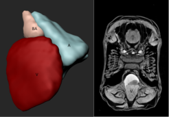
Fig. 11: Microcomputer tomography (µCT) of ex-vivo zebrafish heart. (a) fixed ……..X-ray voltage: 55 KV, X-ray current: 72 µA, filter: Al 0.25 mm, image pixel size: 2.9 µm, tomographic rotation: 180°, rotation step: 0.250°, frame averaging: 4, scan duration: 1h11min. Pictures provided by Anabela Bensimon-Brito, Department III.


