Imaging Platform

Imaging Platform

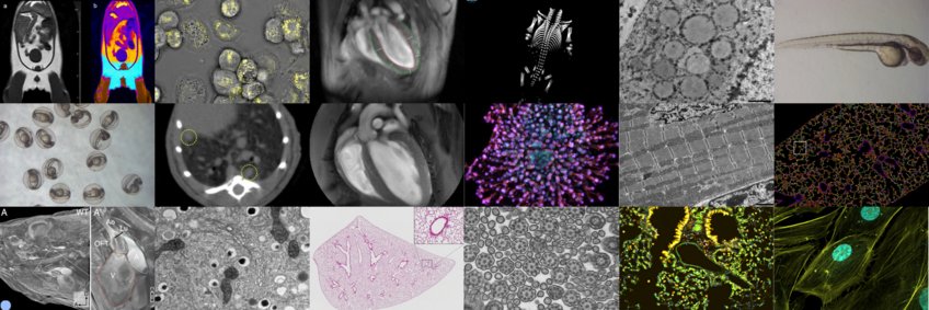
The central imaging core facility at the MPI for heart and lung research, provides a wide range of services and trainings, project specific support and full services for more than 100 scientists at the institute as well as for their collaboration partners. We also offer training for image analysis, develop custom analysis pipelines and help with data storage and archiving.
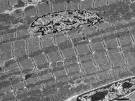
Electron Microscopy
Transmission electron microscopy is available as a full-service including room temperature sample preparation, sectioning, imaging and help with data analysis. Our equipment is well suited for various techniques such as ultrathin sections, negative stainings and CLEM workflows.

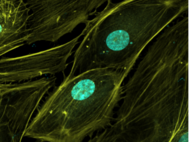
Light Microscopy
We facilitate access to a variety of light microscopes. Most of our equipment can be used as self-service. We provide access to confocal microscopy, 2-photon microscopy, laser microdissection, widefield microscopy and slide scanners. Workflow optimizations and collaborations for complex projects are possible as well.
Magnetic Resonance Imaging (MRI)
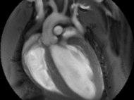
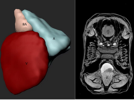
Magnetic Resonance Imaging (MRI) is a non-invasive imaging technique. Without the need for ionizing radiation, it is especially useful for studies that require repeated measurements and need contrast in soft tissues such as liver, brain and heart. Our team and technical setup specialize in small animal cardiac MRI, but we are also equipped for measurements of various other organs. Such as brain, fat and liver.
X-Ray Computed Tomography (µCT)
µCT gives high contrast in areas of different density (examples). Our in vivo scanner is equipped to image living small animals as well as ex vivo samples up to an isometric voxel size of 2.9 µm.
Image Analysis
We provide access to a wide range of open source and commercial tools for the analysis of imaging data through access to several GPU workstations. We also develop custom pipelines for the data analysis from scratch and help with experiment design and data interpretation.
Go to Editor View